Tilly’s Turbulent Time
Sweet little 6-year-old pug Tilly was referred to us at Anderson Moores as she had been experiencing a lack of coordination of her back legs for a few weeks.
She came straight in to see Tomas Elvira, one of our Neurology Residents, who performed a thorough neurological examination. He suspected that Tilly had a lesion affecting the spinal cord in the middle of her back (thoracolumbar region), affecting the messages to and from her back legs.
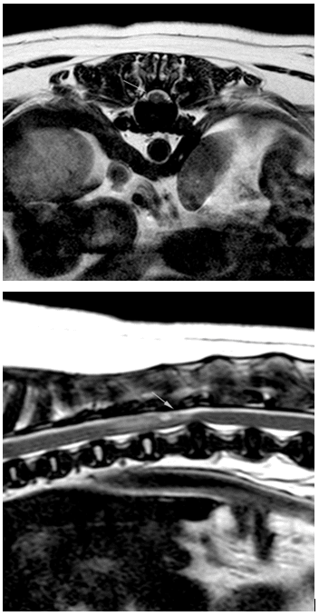
Tilly had a CT and MRI scan performed which revealed an accumulation of fluid around her spinal cord at the level of her last rib (called an ‘arachnoid diverticulum’), together with several misshapen joints between the bones of her spine (hypoplastic articular processes).
Miguel Benito, one of our Neurologists, with Tomas assisting performed a complex surgical procedure to manage her problem. They first created a window in the top of Tilly’s spine to access the spinal cord, before Miguel opened the thin layers that cover the spinal cord (meninges) to surgically release this abnormal collection of fluid and reduce the pressure on the spinal cord. He also broke down all the small adhesions between the spinal cord and the membranes to reduce the chance of it reforming. Following this, Miguel and Tomas used custom-made 3D-printed guides to stabilise Tilly’s spine in this region using screws and bone cement.
Tilly then recovered in hospital for two days under the care of our nursing team day and night.
Tilly’s movement has already improved since she was discharged from hospital. Two weeks after her surgery with us in Hampshire, she came back to be checked and her owner was so happy with her progress and the difference it has made to her movement.
