Teddy’s heart troubles
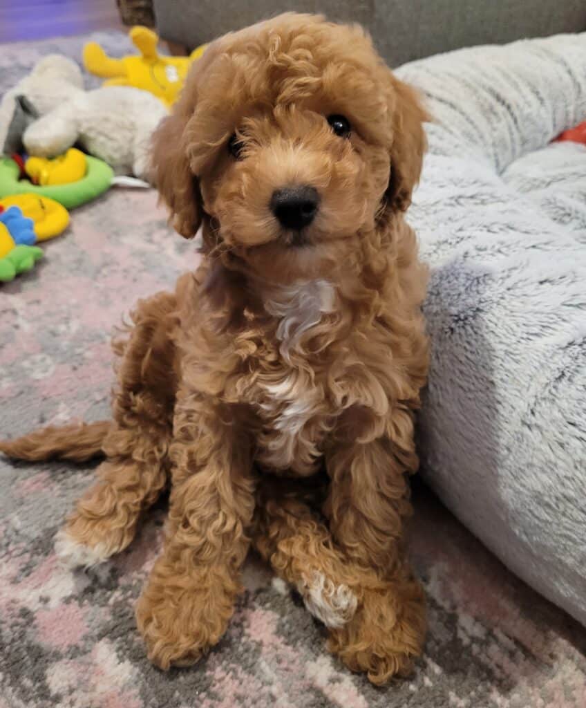
At only 6 weeks old sweet little cockerpoo Teddy visited her vet as she wasn’t well and was told she had a heart murmur so was referred to Valentina Palermo our Head of Cardiology. After a number of tests, she was diagnosed with two congenital heart defects; patent ductus arteriosus (PDA) and severe pulmonic stenosis that are common conditions in dogs.
In patent ductus arteriosus (PDA) the ductus (hole) that is needed during the fetal stage of development in the womb to ensure oxygenation of the fetal blood doesn’t close after birth. Basically, the puppy’s lungs expand with their breathing and the pulmonary vasculature becomes active so this usually closes the ductus.

In some puppy’s, the ductus remains open, making an abnormal link or ‘shunt’ between the dog’s aorta and their pulmonary artery. If pressures remains, then the blood that shunts from the aorta to the pulmonary artery causes over-circulation and dilatation of the left side of their heart. If left untreated, dogs with PDAs ultimately develop left-sided congestive heart failure (CHF). The treatment required is therefore to close the PDA.

This is performed by surgery either surgical ligation where the surgeon opens up the dog’s thorax and ligates the ductus with sutures – so ties them off. This technique is invasive with a more prolonged recovery. However, it allows the surgeon to be able to see the ductus more clearly in very small puppies. There is also an option for minimally invasive surgery where there is a much quicker recovery time for them.
Teddy was also diagnosed with a severe form of pulmonic stenosis. The severe nature of her right ventricular outflow tract obstruction meant that she had right ventricular thickening and post-stenotic dilatation of her main pulmonary artery. Her valves were thickened and fused and therefore we knew Teddy would benefit from balloon valvuloplasty. With this minimally invasive procedure, a balloon catheter is placed into the main pulmonary artery, across the pulmonic valve. With the inflation of the balloon, the valve opens relieving the obstruction.


In February 2022 Teddy underwent general anaesthesia for this surgery. The first procedure Valentia and the team performed was PDA closure by implanting a device, which blocks the hole and prevents blood from shunting across it. The procedure was successful, so they then proceeded with the second procedure, the balloon valvuloplasty to reduce the pulmonic stenosis severity.

The procedure was completed without any complications and Teddy recovered well from the anaesthetic.

A heart scan the next day showed that the implant was safely in place, with no blood flow going through the PDA. The team were delighted as a successful implantation should completely cure a PDA. They were also very happy to notice a marked improvement in Teddy’s pulmonic stenosis, which improved from severe to mild, and if this remains mild, Teddy will not require medication, should not develop any clinical signs and live a normal life. A real success for the team on such a tiny young patient.

