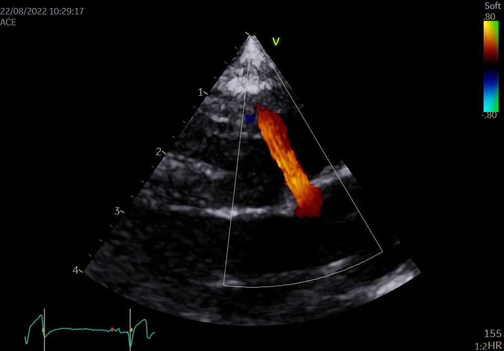
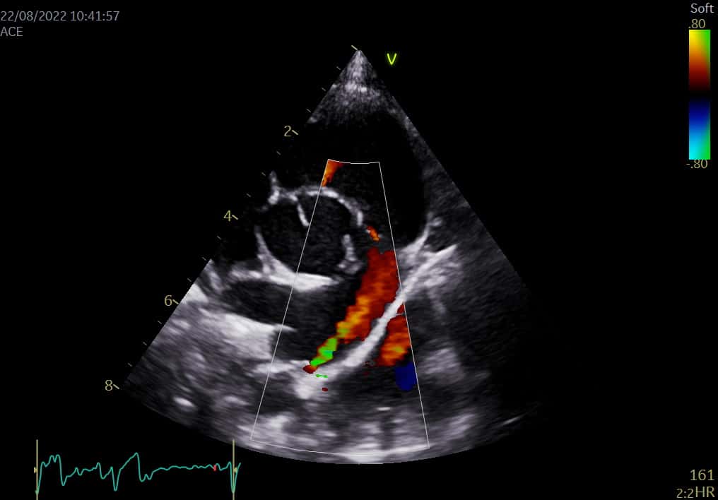
Sweet little 5-month-old wire hair fox terrier Pike was referred to Valentina Palermo our head of Cardiology over a concern with his heart. She diagnosed him with a small Ventricular Septal Defect (VSD). A VSD is a hole between the two ventricles in the heart.

A dog’s heart is a muscular pump with four separate chambers. The right side of the heart sends blood to the lungs where it picks up oxygen. The left side of the heart pumps the blood around the body. The heart is divided into left and right by a muscular wall (the septum). The ventricular septum separates the right and left ventricle.
In a VSD the wall doesn’t develop properly resulting in a small ‘hole’ in the septum allowing some blood to pass from the left side of the heart to the right side. The effects of this depend on the size and location of the defect.

In some cases, very small VSD holes may close spontaneously. Larger defects can lead to congestive heart failure (fluid within the lungs).
Pike had a small VSD and his heart measured normal, so it is possible that this defect could close with time or remain very small with a minimal amount of blood passing through.
Pike was also diagnosed with Patent Ductus Arteriosus (PDA), one of the more common congenital heart defects in dogs. A PDA is caused by a foetal vessel (the ductus arteriosus) failing to close at birth. The ductus carries blood away from the foetus’ lungs but should close at birth. If it remains open some blood that should travel from the left heart to the body, via the aorta, is diverted into the pulmonary artery and travels to the lungs.

The long-term consequence of this is increased blood flow through the left heart and lungs, which causes the left heart to stretch and dilate and can eventually lead to Congestive Heart Failure (CHF) which is when the heart is unable to cope with the excess flow and fluid builds up inside the lungs. Most dogs with PDAs will develop CHF at a young age, and many die before they are two years old, so closure is highly recommended in these patients.
PDAs can either be closed by a minimally invasive catheter-based procedure or by standard surgery. As Pike’s heart was normal size, we have decided to assess him again in three to six months to assess any progression and decide if he requires surgery to close the PDA.